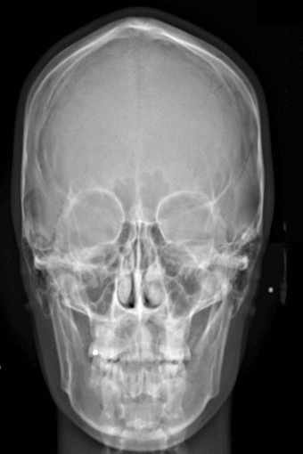
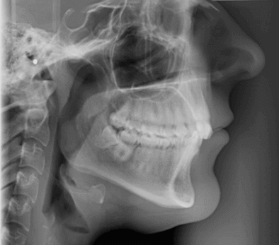
CEPH X-Ray
A Cephalometric x-ray (CEPH x-ray) consists of a lateral/side-view x-ray of the face - also referred to as Lat CEPH. As with an OPG X-ray a CEPH radiograph is used to study facial bone and soft tissue landmarks and is used in patients being considered for orthodontic treatment.
For the CEPH x-ray, an adapted panoramic OPG x-ray machine is used. The patient is in the standing position and the cephalometric radiograph of the head is taken using a cephalometer (Cephalostat) that holds the head in the correct position in readiness for the CEPH x-ray.
Digital CEPH X-rays
Once a CEPH x-ray is taken a computerised cephalometric process converts the captured image into a digital format. The CEPH x-ray in digital format allows for zooming into area of interest, such as potential TMJ problems, and/or impacted/ non-erupted dentition. The digital x-ray also has the advantage that it allows for measurements of teeth and bone and enables annotations to be made directly on the digital CEPH image.
CT Dent has taken the digital CEPH a step further incorporating digital cephalometric tracing with Artificial intelligence (AI) software – See the links HERE for more information.
Click on the image below to open up an example of a digital CEPH x-ray image - The image is shown using the PACS Cloud Viewer and allows you to take measurement, add annotations and use the zoom functionality.
Panoramic Digital Cephalometric
A panoramic digital cephalometric (PA Digital CEPH) consists of a radiograph of the head taken with the x-ray beam perpendicular to the patient’s coronal plane with the x-ray source behind the head and the film cassette in front of the patient’s face.
OPG x-rays
Click link HERE for information on OPG x-rays.
Quick query
Call Us Today+44 (0)20 7487 5717


