

Cone Beam CT (CBCT)
Cone Beam CT Scan
Dental Cone Beam CT scans (CBCT Scans) provide high resolution, 3D volumetric images, used in Dental Implant Planning, Orthodontics and Maxillofacial surgery. allowing for more accurate analysis of bone structure and dental/tooth orientation. The accuracy of a CBCT scan is comparable to medical CT scans but uses a much lower radiation dose but with the added advantage of greater accuracy of bone structures and dental/tooth orientation. Your patient sits in a chair, as opposed to lying in a tunnel, and the time for a dental CBCT scan is just twenty seconds.
Whereas typical dental X-rays focus on a small area to produce flat, 2D image, CBCT scan images enable a more complete and accurate 3D images - allowing you to more effectively plan patient surgical and dental treatments. Individual patient Cone Beam CT images are also digitally stored for ease of access in the future, and for sharing with colleagues or patients.
Cone Beam CT scans can be used in multiple situations and offers benefits to you and your patients across the following areas:
Dental Implants:
The high quality of CBCT scans allows you to more accurately plan and place dental implants - from initial stages of diagnosis through to treatment and post-operative examinations.
Orthodontics:
CBCT in orthodontics provides clear and accurate images for dental brace placement. Orthodontic CBCT provides accurate tooth relationships and assessment of dental asymmetry.
Impacted Teeth:
A CBCT scan can highlighted whether a patient has impacted teeth and accurately determine their precise position in relation to adjacent teeth and associated roots, and proximity to important structures such as nerve canals, sinus walls and cortical borders.
Patient Airway Assessment:
Restricted airways are susceptible to collapse but are not always easy to pinpoint. Cone beam CT scan of a patients’ airway allows you to reconsider your treatment plan if a problem is found.
Temporomandibular Joint (TMJ:
Temporomandibular joint CBCT (TMJ CBCT) is not only cost-effective but allows complete TMJ analysis and diagnosis of multiple TMJ conditions, providing clear images of joint spacing and any dysfunction of condyles and surrounding structures.
Analysis:
Cone Beam CT scan analysis can be used in detecting and evaluating possible or unforeseen patient problems for your patients, thanks to accurate measurements of bone and jaw deformities. This allows you to assess bone lesions and changes of the jaw and detect cysts, tumours, and disease.
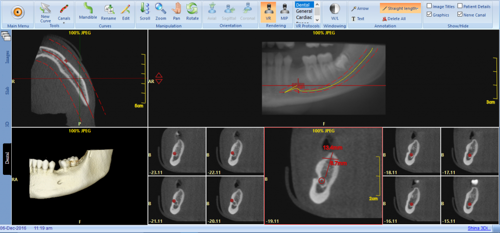
CBCT Working Example:
Click on the image below to open up a working example of a Cone Beam CT scan visualised using the PACS Cloud Viewer
Quick query
Call Us Today+44 (0)20 7487 5717