

Cases

CT Dent opens 7th UK dental imaging centre in Colchester
July 17, 2018
CBCT IR(ME)R referrer training – Register for our forthcoming course
October 2, 2018Case of the Month – Impacted Teeth
CBCT Scanner: i-CAT Classic
CBCT Imaging Protocol: 60x80x80mm, 0.2 voxel
Effective Dose: 0.03 mSv
Clinical Information: Assessment of location of premolars and molars and relationship with IDN
Radiographic Impression:
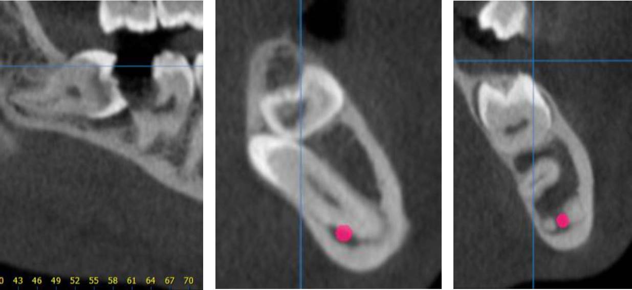
Mandible: The right mandibular premolars and molars are impacted. The primary second molar is ankylosed and impacted; it seems to be preventing the eruption of 35 and directing it in a lingual position. The mesial aspect of the crown of 36 is in contact with the impacted primary tooth and presents with an external resorption.
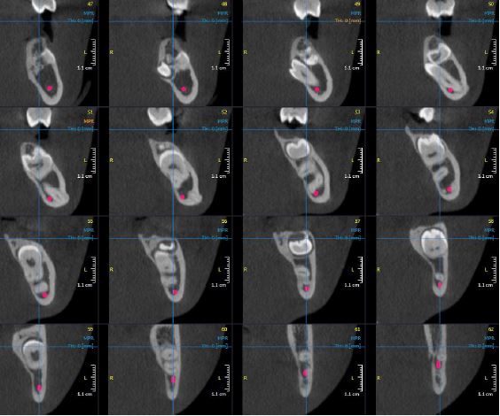
36, 37 and 38 present with apical dilaceration, the apices of 38 are situated within the mandibular canal.
Impressions and recommendations:
Ankylosed primary tooth with impaction of 35, 36, 37 and 38.
35 is in lingual position, 36-38 are impacted and their roots are dilacerated.
48 impacted in an oblique direction with large carious lesion on 47.
CBCT Imaging Protocol: 60x80x80mm, 0.2 voxel
Effective Dose: 0.03 mSv
Clinical Information: Assessment of location of premolars and molars and relationship with IDN
Radiographic Impression:
Mandible: The right mandibular premolars and molars are impacted. The primary second molar is ankylosed and impacted; it seems to be preventing the eruption of 35 and directing it in a lingual position. The mesial aspect of the crown of 36 is in contact with the impacted primary tooth and presents with an external resorption.
36, 37 and 38 present with apical dilaceration, the apices of 38 are situated within the mandibular canal.
Impressions and recommendations:
Ankylosed primary tooth with impaction of 35, 36, 37 and 38.
35 is in lingual position, 36-38 are impacted and their roots are dilacerated.
48 impacted in an oblique direction with large carious lesion on 47.
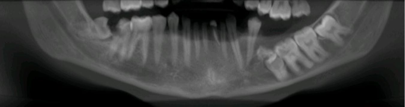
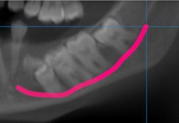
Panoramic reconstruction
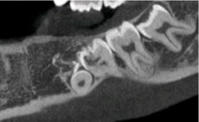
External resorption on 36
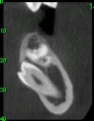
35
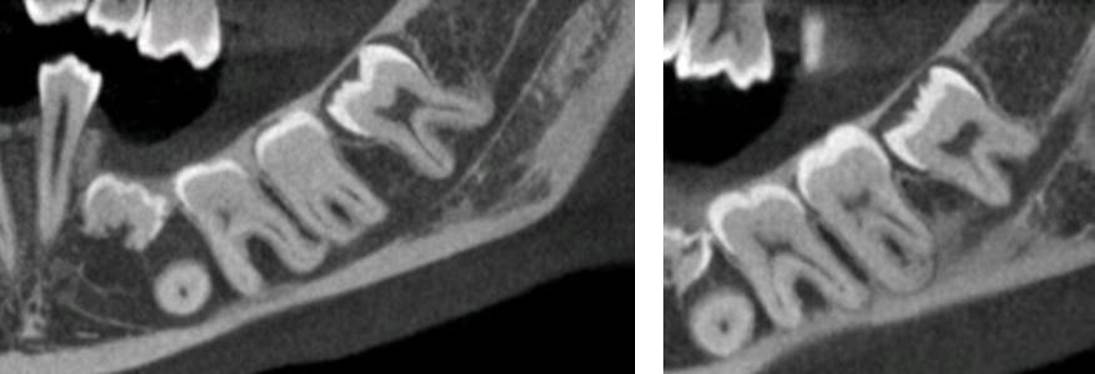
Dilacerated roots
47-48
Left IDN highlighted
Radiographic Impression:
Mandible: The right mandibular premolars and molars are impacted. The primary second molar is ankylosed and impacted; it seems to be preventing the eruption of 35 and directing it in a lingual position.