

Cases

CT Dent British Dental Journal Article
June 28, 2018Case of the month – Patient Experiencing Paraesthesia
CBCT Imaging Protocol: 60x60x80mm, 0.2 voxel
Effective Dose: 0.06mSv Clinical Information: Patient experiencing paraesthesia in lower right quadrant following implant placement.
Radiographic Impression
Reconstructed panoramic
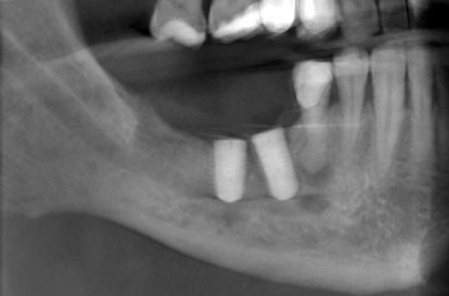
Mandibular right premolar-molar region
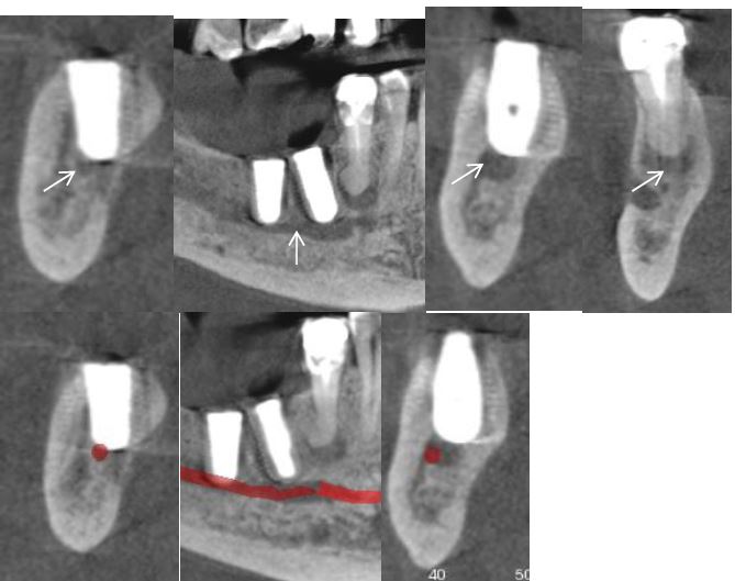
The small arrows illustrate the mandibular canal; both implants appear to interrupt/extend into the cortical boundary separating the alveolar process from the mandibular canal. A well circumscribed periapical radiolucency was observed associated with the apex of the mandibular right second premolar; the apex of tooth also exhibits hypercementosis.
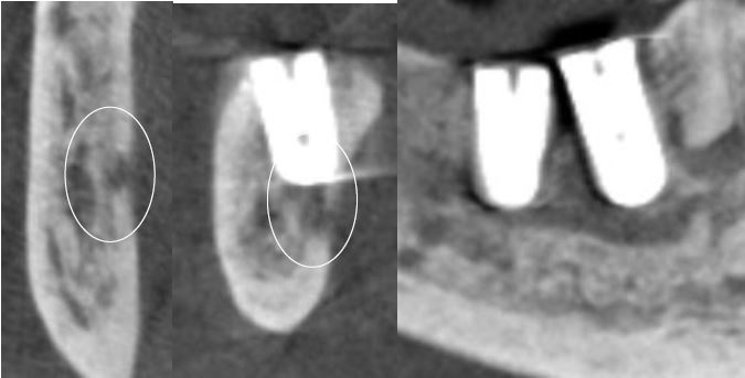
The more posterior mandibular implant exhibits a localized irregular area of low density lingual and inferior to the apex of the implant [circle] that appears to thin and potentially perforate the lingual cortical plate; the area may potentially represent osteotome penetration or localized inflammatory change. Careful clinical evaluation of the area is suggested to rule out a localized area of osteomyelitis.
Our latest case of the month: Patient experiencing paraesthesia in lower right quadrant following implant placement.