Cases


The Importance of CBCT in Implant Dentistry
March 12, 2019

The development of artificial intelligence applications at CT Dent – working with Orca Dental AI
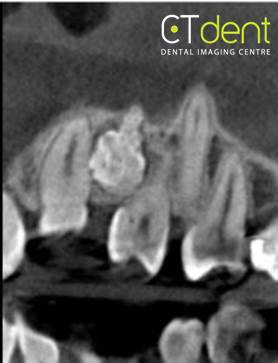
April 9, 2019Case of the month – A Case of a Compound Odontoma
CBCT Imaging Protocol: 60cm x 40cm x 40cm, 0.13 voxels
Effective Dose: 0.06 mSv
Clinical Information: Radiopacity seen in upper left anterior region. Confirmation required of likely diagnosis and relationship with adjacent teeth.
Click here to view and manipulate this case of the month CBCT on our Cloud Viewer
Radiographic Impression:
Dental Findings: Specific comments associated with each of the cross-sections below.
The following are selected images from the volume illustrating major findings
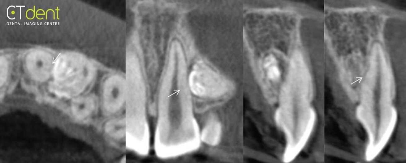
Maxillary left central incisor region; the cross-sections are oriented with the long axis of the central incisor. The central incisor exhibits a normal crown root ratio. The compound odontoma is positioned in close physical proximity to the distal lingual surface, mid root of the central incisor. Localised areas of mild to moderate resorptive change were noted [arrows].
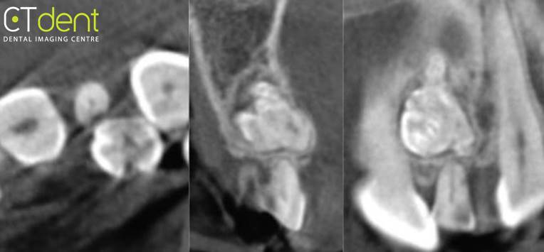
Maxillary left primary lateral incisor; the apex of the primary lateral incisor exhibits moderate to severe resorption. The odontoma appears positioned superior to the apex of the primary lateral incisor and centrally positioned within the alveolar process.

Maxillary left lateral incisor; the tooth exhibits multiple coronal structural abnormalities [arrows]; the root of the tooth is curved labially. The tooth is positioned on the lingual surface of the alveolar process palatal to the odontoma. The PDL space appears to be intact with no radiographic evidence of ankylosis. The small circle illustrates a portion of the curved root apex.
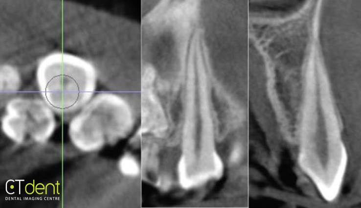
Maxillary left canine; cross-sections are oriented with the long axis of the canine. The tooth exhibits a normal crown root ratio with no radiographic evidence of root resorption.
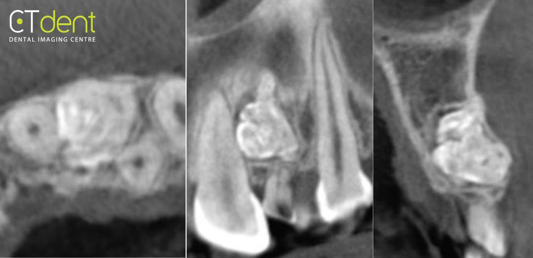
The compound odontoma is positioned centrally within the alveolar process distal to the central incisor and labial to the adult lateral incisor. The odontoma exhibits an irregular surface contour that extends labially, thinning and potentially perforating the labial cortical plate.
Clinical Information: Radiopacity seen in upper left anterior region. Confirmation required of likely diagnosis and relationship with adjacent teeth.


