

Cases

COVID-19 update from CT Dent
March 20, 2020
Dental imaging that enhances your clinic
November 18, 2020Case of the Month – Implant Planning; Accessory Neurovascular Canal
CBCT Scanner: Instrumentarium OP300
Scanning Protocol: 8cm x 6cm FOV; 0.2 voxels
Effective Dose: 0.08 mSv
Clinical Information: Assessment of the position of anatomical structures and pathology prior to implant placement in the lower left molar region. Patient wearing radiographic template.
Click here to view and manipulate this case of the month CBCT on our Cloud Viewer
Radiographic Impression: A small accessory vascular canal is seen branching from the left mandibular canal. Bone volume measurements have been provided for the marked sites of the radiographic template.
The following are selected images from the volume illustrating major findings
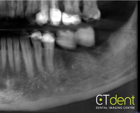
Reconstructed panoramic radiograph
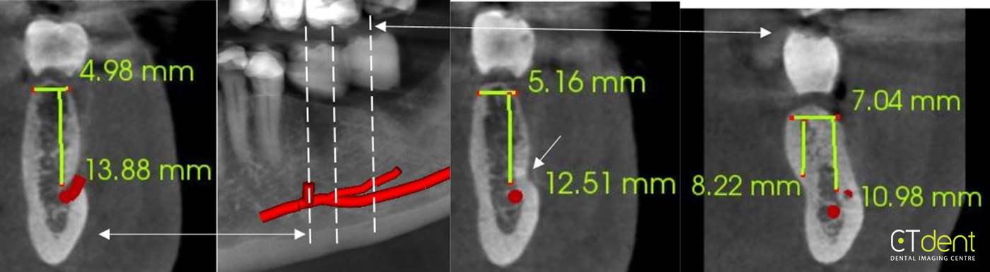
Cross-sections through the edentulous mandibular left molar region; a small accessory vascular canal branches from the primary mandibular canal extending buccally and posteriorly.
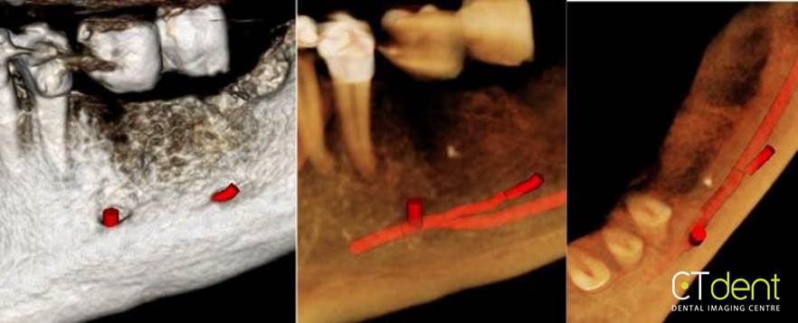
3D rendering of the accessory vascular canal
Clinical Information: Assessment of the position of anatomical structures and pathology prior to implant placement in the lower left molar region. Patient wearing radiographic template.