

Cases

Past, Present and Future CBCT
January 14, 2019
Dr Nilesh Parmar discusses his experience of working with CT Dent
February 12, 2019Case of the Month – Planning for Dental Implants
CBCT Scanner: KaVo OP 3D Vision V17
CBCT Imaging Protocol: 16cm height x 10cm width, 0.25mm voxel
Effective Dose: 0.15 mSv
Clinical Information: Scan assessment prior to implants in mandible and zygoma.
Click here to view and manipulate this case of the month CBCT on our Cloud Viewer
Main Findings:
Maxilla: No abnormalities detected
Sinuses: A generalised increase in the thickness and density of the tissues lining the right and left maxillary, ethmoid, and sphenoid sinuses were noted. The bone forming the maxillary and sphenoid sinuses is thickened and sclerotic/increased in density.
Nasal Cavity: No abnormalities detected
Mandible: No abnormalities detected
Air Space: Mild enlargement of the pharyngeal tonsils was noted.
TMJs: No abnormalities detected; visible portions of the condyle exhibit a smooth continuously corticated outline and good symmetry.
Other findings: Curvilinear areas of increased density were noted lateral to the pituitary fossa in areas anatomically associated with the carotid arteries; these areas appear to be consistent with calcification of the carotid artery.
Dental findings: no abnormalities detected
Radiographic Impression:
Sinuses: The radiographic findings are consistent with a generalised, long-standing chronic mucositis/sinusitis of the right and left maxillary sinuses. The thickened, sclerotic walls of the maxillary and sphenoid sinuses are an indication of long-standing chronic inflammatory changes. Correlation of the radiographic observation with the patient's clinical history and symptoms if any is suggested. Physician referral for more thorough evaluation is suggested.
Airspace: Clinical evaluation of the soft tissues of the oropharynx is suggested.
Other Findings: Vascular calcifications may be an indication of a potential increased risk of cardiovascular disease and stroke. Correlation of the radiographic observation with the patient’s clinical history of high blood pressure, elevated cholesterol, smoking and stress is suggested. Physician referral for more thorough evaluation is suggested if merited by clinical findings.
The following are selected images from the volume illustrating major findings
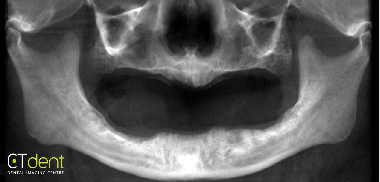
Reconstructed panoramic radiograph
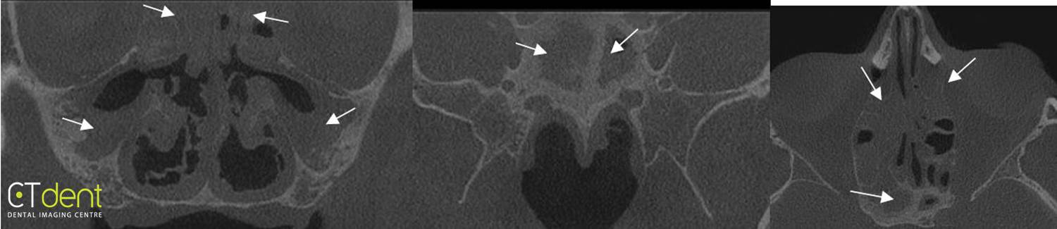
A generalized increase in the thickness and density of the tissues lining the right and left maxillary, ethmoid, and sphenoid sinuses were noted. The bone forming the maxillary and sphenoid sinuses is thickened and sclerotic/increased in density.
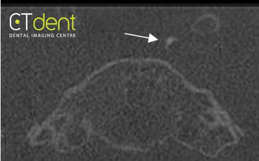
Curvilinear areas of increased density were noted lateral to the pituitary fossa in areas anatomically associated with the carotid arteries; these areas appear to be consistent with calcification of the carotid artery.
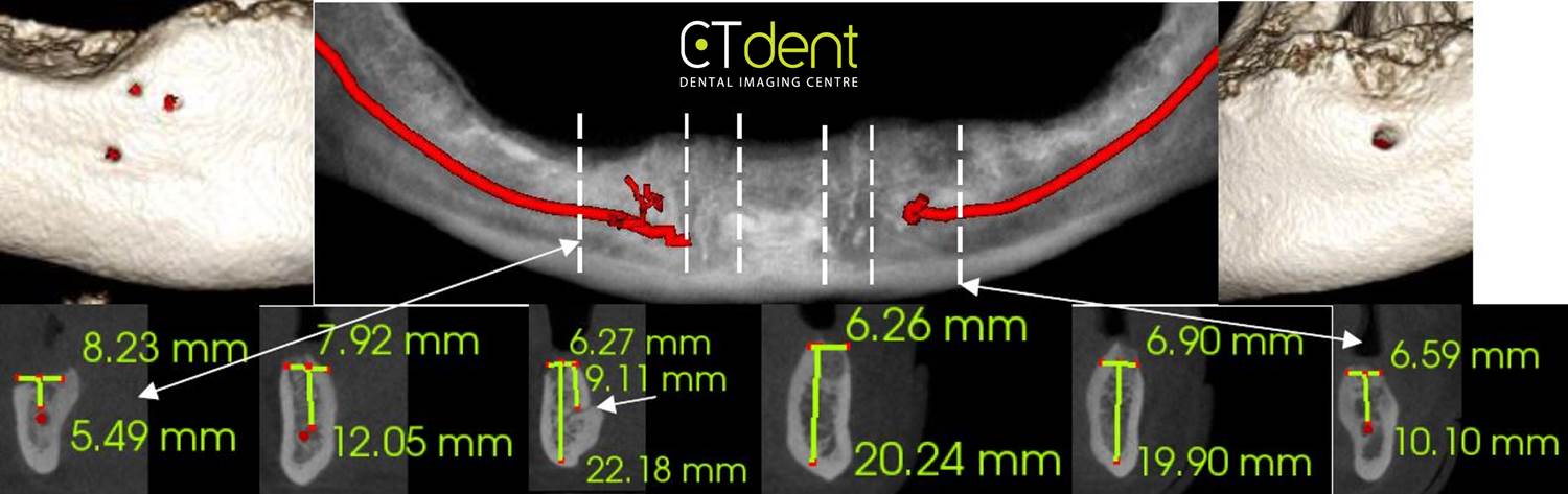
Cross-sections through selected areas of the mandible; the small arrow illustrates the lingual foramen and canal. The mental foramen on the right is formed by three small vascular branches off of the mandibular canal whereas the left mental foramen exhibits a more traditional appearance.
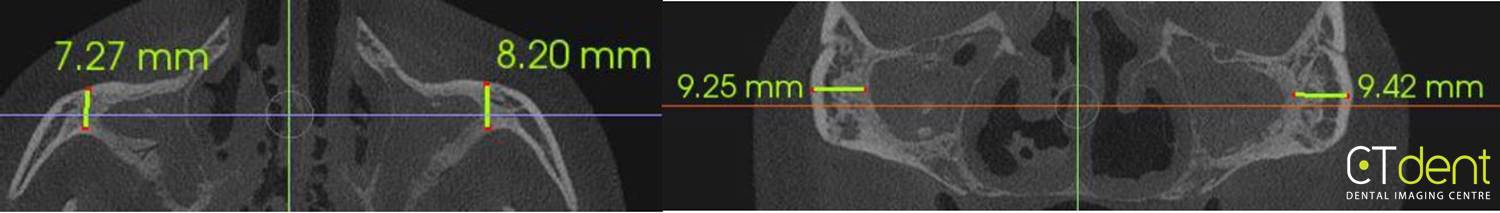
Cross-sections through selected areas of the maxilla
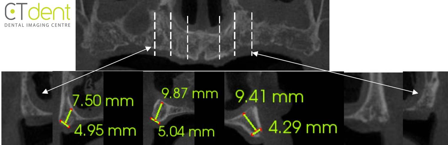
Measurements through selected areas of the zygomatic arch
Main Findings:
Maxilla: No abnormalities detected
Sinuses: A generalised increase in the thickness and density of the tissues lining the right and left maxillary, ethmoid, and sphenoid sinuses were noted. The bone forming the maxillary and sphenoid sinuses is thickened and sclerotic/increased in density.