

Cases

CBCT IR(ME)R referrer training – Register for our forthcoming course
October 2, 2018CT Dent has been shortlisted as a finalist in the Dental Industry Awards 2018
October 22, 2018Case of the Month – Periapical Lesions
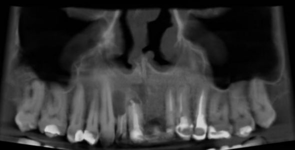
CBCT Scanner: Instrumentarium OP300
CBCT Imaging Protocol: 60x60x80mm, 0.2 voxel
Effective Dose: 0.04 mSv
Clinical Information: Implants planned in upper aesthetic zone
Findings: Paranasal Sinuses: minimal mucosal thickening noted in the right maxillary sinus with some calcifications. A small mucous attention cyst is noted on the floor of the left sinus.
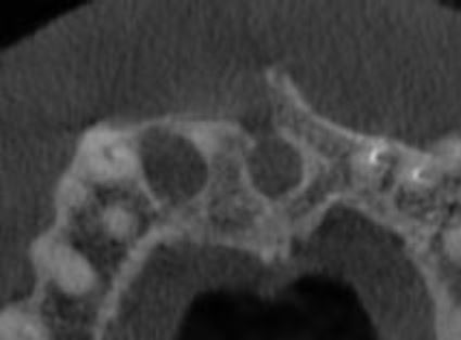
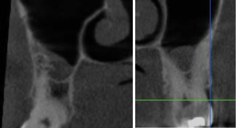
Maxilla: presence of a periapical lesion on 12 with severe external resorption. The lesion is causing thinning on both buccal and palatal cortices. The crown to root ratio of the tooth is unfavorable.
Periapical lesion with endodontic surgery and retrograde obturation are noted on 11 with presence of a bone defect superior to the tooth extending posteriorly to communicate with the incisive canal.
Impressions and recommendations:
Prognosis of 12 is poor with need for extraction, enucleation of the cyst and biopsy.
Prognosis of 11 is also noted with presence of a periapical lesion and another most probably inflammatory lesion involving the incisive canal. Extraction of the tooth and enucleation of both lesions and biopsy is recommended as well.
Mucous retention cysts and mucositis in the maxillary sinuses are common findings, the calcifications are consistent with antrolith, no further evaluation needed.
CBCT Imaging Protocol: 60x60x80mm, 0.2 voxel
Effective Dose: 0.04 mSv
Clinical Information: Implants planned in upper aesthetic zone
Findings: Paranasal Sinuses: minimal mucosal thickening noted in the right maxillary sinus with some calcifications. A small mucous attention cyst is noted on the floor of the left sinus.
Maxilla: presence of a periapical lesion on 12 with severe external resorption. The lesion is causing thinning on both buccal and palatal cortices. The crown to root ratio of the tooth is unfavorable.
Periapical lesion with endodontic surgery and retrograde obturation are noted on 11 with presence of a bone defect superior to the tooth extending posteriorly to communicate with the incisive canal.
Impressions and recommendations:
Prognosis of 12 is poor with need for extraction, enucleation of the cyst and biopsy.
Prognosis of 11 is also noted with presence of a periapical lesion and another most probably inflammatory lesion involving the incisive canal. Extraction of the tooth and enucleation of both lesions and biopsy is recommended as well.
Mucous retention cysts and mucositis in the maxillary sinuses are common findings, the calcifications are consistent with antrolith, no further evaluation needed.
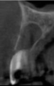
12
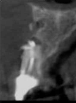
11
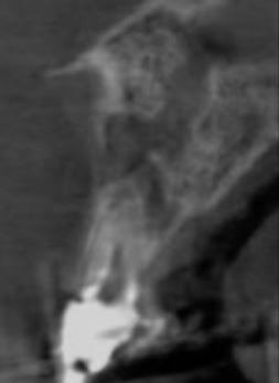
Lesion involving the incisive canal
Anterior maxilla
Maxillary sinuses
Findings:
Paranasal Sinuses: minimal mucosal thickening noted in the right maxillary sinus with some calcifications.
A small mucous attention cyst is noted on the floor of the left sinus.