News


The development of artificial intelligence applications at CT Dent – working with Orca Dental AI
April 9, 2019

Case of the month – Diagnosis of a Bony Lump
May 20, 2019Using CBCT for Investigating Lower Third Molars
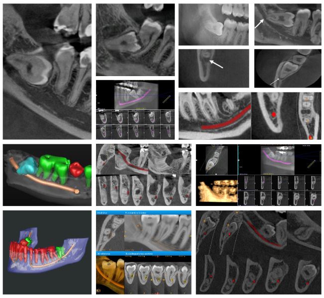
CBCT imaging is used prior to many dental procedures as it allows for a more accurate and safer patient outcome. One such procedure is the extraction of the lower third molar, due to its ability to accurately determine the relationship of the roots of the tooth with the inferior dental canal (IDC).
In many patients, the lower third molar has a very close relationship or is communicating directly with the IDC. Although this can be assessed on periapical and dental panoramic images, using CBCT prior to extraction can significantly reduce the risk of post-operative complications such as paraesthesia.
CBCT can also be used to look at the close relationship of the lower third molar with the second molar and assessing potential crown and root pathology, for example areas of decay and resorption. It can also highlight early follicular changes which can lead to dentigerous cyst development.
CT Dent has been providing an independent dental imaging service to dental practitioners since 2007. All of our centres use the latest in cone beam CT imaging technology, providing high resolution, 3D volumetric images for diagnostic analysis and treatment planning.
Our constant investment in new technology means those using our service receive consistently high-quality scans. All our X-ray imaging at CT Dent is taken by diagnostic radiographers who have previously worked in hospitals and have chosen to specialise in dental and maxillofacial radiography.
REGISTER WITH CT DENT

CBCT imaging is used prior to many dental procedures as it allows for a more accurate and safer patient outcome, one such procedure is the extraction of the lower third molar.


