

Cases
Case of the month – Identifying expended lesion
July 16, 2021
Our New AI + Human Radiology Reports are here!
February 6, 2024Case of the month – Implant analysis requested. Pre-implant UL1
CBCT Scanner: Kavo OP300
Scanning Protocol: 5cm x 5cm FOV; 0.12 voxels
Effective Dose: 0.04 mSv
Clinical information: Implant analysis requested. Pre-implant UL1.
Scanning Protocol: 5cm x 5cm FOV; 0.12 voxels
Effective Dose: 0.04 mSv
Clinical information: Implant analysis requested. Pre-implant UL1.
Radiographic Impression
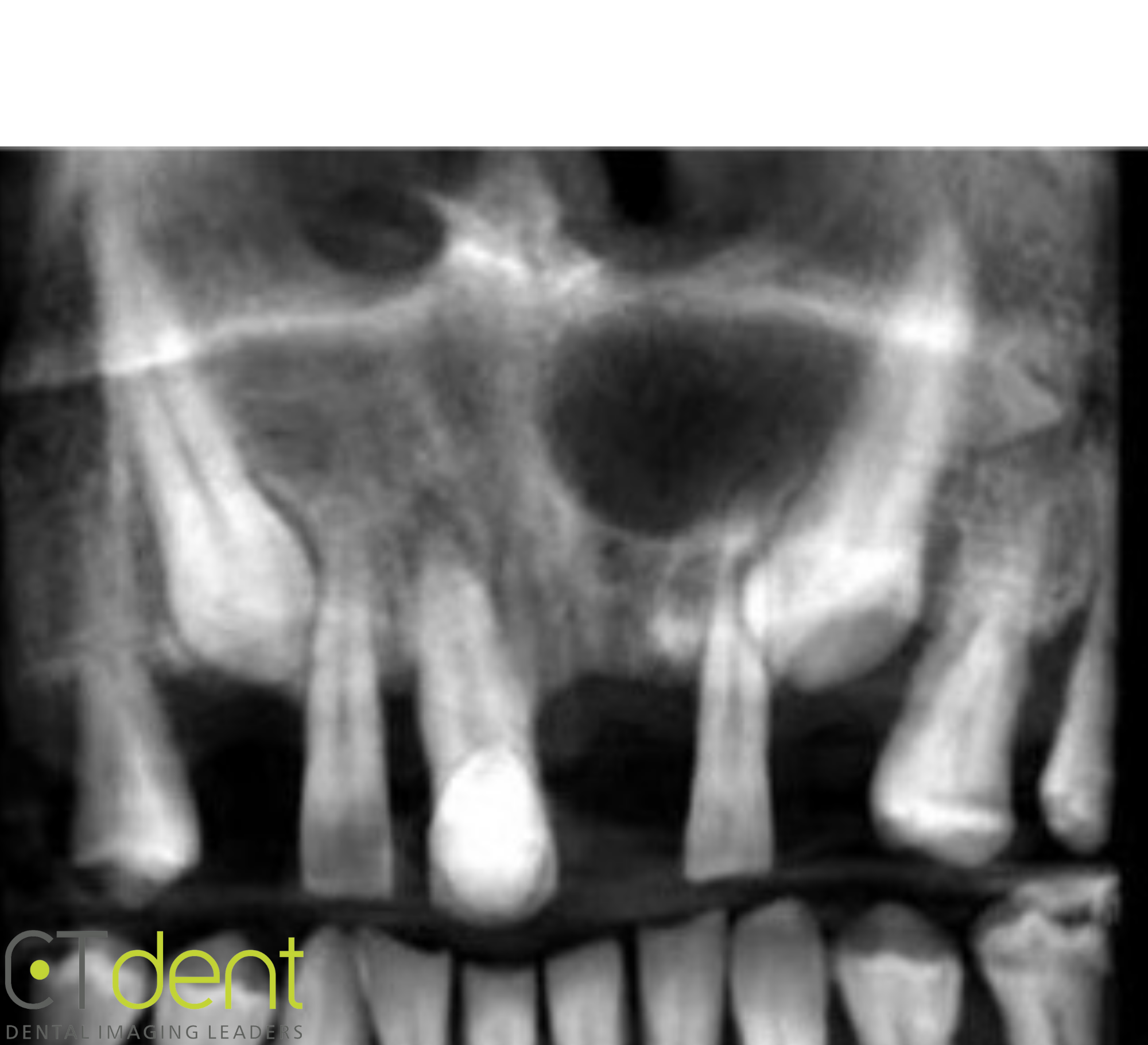
Reconstructed panoramic radiograph
Dental Findings:
Maxillary left central incisor region; a small supernumerary tooth exhibiting internal/external resorptive changes was observed on the distal surface of the alveolar process with the crown adjacent to the lingual surface of the canine-first premolar interproximal space. The area also contains a large well circumscribed area of low density/radiolucency suggestive of a previous periapical radiolucency/low density. Correlation of the radiographic observation with the patient’s clinical/surgical history is suggested.
Radiographic Impression:
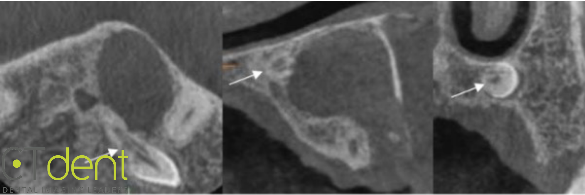
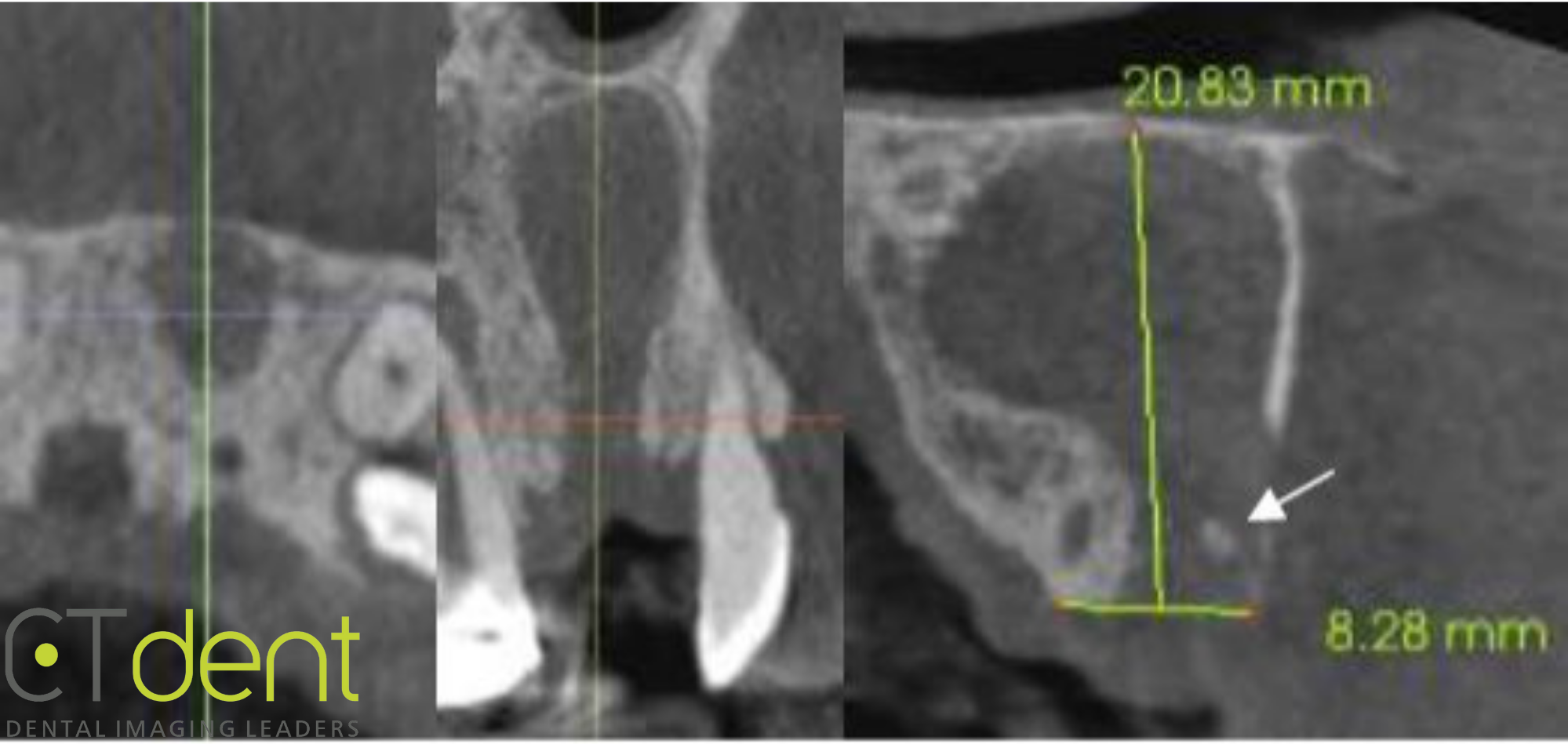
Maxillary left central incisor region. The labial cortical plate appears to be very thin are absent. In addition, a small ovoid area of increased density was noted within the extraction socket suggestive of a small residual root/bone fragment [arrow].
Maxillary right central incisor. A small well circumscribed periapical radiolucency/low density was observed associated with the apex of the tooth. The radiographic appearance is consistent with periapical inflammatory disease of pulpal origin. Appropriate clinical evaluation is suggested.
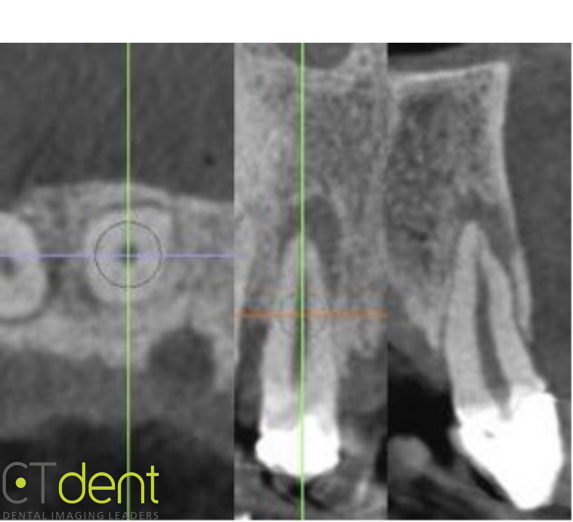
Click here to view and manipulate this case of the month CBCT on our Cloud Viewer
The following are selected images from the volume illustrating major findings
Click here to view and manipulate this case of the month CBCT on our Cloud Viewer
The following are selected images from the volume illustrating major findings
Maxillary left central incisor region; a small supernumerary tooth exhibiting internal/external resorptive changes was observed on the distal surface of the alveolar process with the crown adjacent to the lingual surface of the canine-first premolar interproximal space.