

Cases

Case of the month – Diagnosis of a Bony Lump
May 20, 2019
CBCT Level 1 training – ONLINE COURSE
July 16, 2019Case of the Month – All-on-Four and Zygomatic Implant Planning
CBCT Imaging Protocol: 14cm diameter x 8.5cm height, 0.25mm voxel
Effective Dose: 0.095 mSv
Clinical Information: Pre-implant assessment for all-on-4 procedure, and potential planning for zygomatic implants if necessary.
Radiographic Findings:
A small dome-shaped homogeneous area of increased density was observed in the sphenoid sinus. Periapical radiolucencies were associated with the maxillary right canine and maxillary left first and second premolars. Curvilinear areas of increased density were noted lateral to the pituitary fossa in areas anatomically associated with the carotid artery; these appears to be consistent with calcification of the carotid artery. Such calcifications may be a potential indication of an increased risk of cardiovascular disease and stroke. Correlation of the radiographic observation with any clinical history of high blood pressure, elevated cholesterol, smoking and stress would be suggested. The left external auditory meatus appears partially obstructed by an irregular area of increased density consistent with earwax accumulation, which may diminish hearing acuity; correlation of the radiographic observation with the patient’s clinical history is suggested.
The following are selected images from the volume illustrating the findings
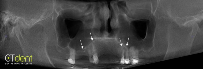
Reconstructed panoramic radiograph; a localised area of increased density was observed in the maxillary right second premolar region and appears consistent with residual dental material. Periapical radiolucencies were associated with the maxillary right canine and maxillary left first and second premolars.
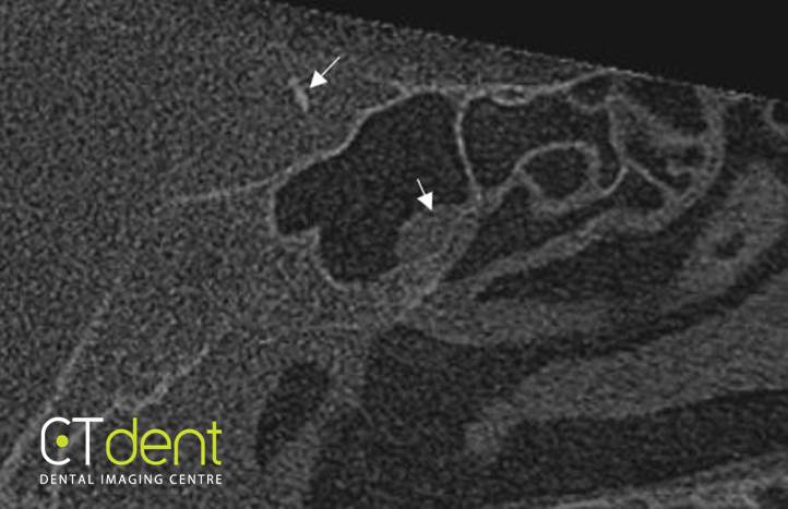
Curvilinear areas of increased density were noted lateral to the pituitary fossain areas anatomically associated with the carotid artery; the area appears to be consistent with calcification of the carotid artery. A small dome-shaped area of increased density was observed within the sphenoid sinus. The right and left osteomeatal complexes were patent.
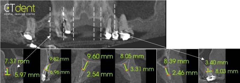
Cross-sections through selected areas of the maxilla.
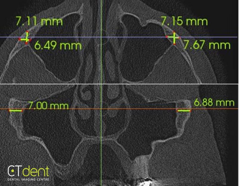
Cross-sections through the zygomatic arch.
Clinical Information: Pre-implant assessment for all-on-4 procedure, and potential planning for zygomatic implants if necessary.