

Cases

Our AI is Learning Fast
October 2, 2019
Case of the month – Implant Planning
November 27, 2019Case of the Month – Dentoalveolar Trauma
CBCT Scanner: KaVo OP300 Instrumentarium
CBCT Imaging Protocol: 5cm diameter x 5cm height, 0.125mm voxel
Effective Dose: 0.037 mSv
Clinical Information: Patient was hit in the face with a squash racket. UR1 horizontal fracture seen on periapical. CBCT needed to assess extent and any associated bony pathology.
Click here to view and manipulate this case of the month CBCT on our Cloud Viewer
Findings:
Maxilla: No abnormalities detected
Sinuses: No abnormalities detected
Nasal Cavity: No abnormalities detected
Other findings: No abnormalities detected
Dental findings: Horizontal fracture of the maxillary right central incisor; the remainder of the teeth included in the volume appear to be normal.
Radiographic Impression:
Dental Findings: Horizontal fracture of the maxillary right central incisor.
The following are selected images from the volume illustrating major findings
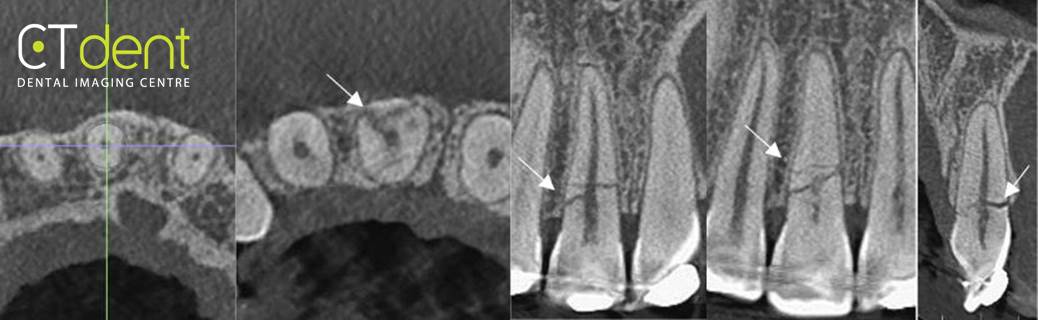
Maxillary right central incisor. The tooth exhibits a horizontal fracture just apical to the alveolar crest. The labial and lingual cortical plates appear to be intact; radiographic evidence of possible fracture of the alveolar process was not identified.
Clinical Information: Patient was hit in the face with a squash racket. UR1 horizontal fracture seen on periapical. CBCT needed to assess extent and any associated bony pathology.