

Cases

Case of the Month – Radicular cyst
November 25, 2020
Patient safety is our priority
January 5, 2021Case of the Month – Impacted LL3 with focal osseous dysplasia and compound odontoma
CBCT Scanner: Gendex DP700
Scanning Protocol: 6cm x 4cm FOV; 0.13 voxels
Effective Dose: 0.03 mSV
Clinical Information: Orthodontic assessment. Ectopic LL3 positioned near midline. Retained LLC.
Click here to view and manipulate this case of the month CBCT on our Cloud Viewer
Radiographic Impression:
The 33 is impacted in a vertical direction, with the crown thinning and possibly interrupting the lingual cortex. The crown is in contact with the lingual aspect of the 32 and 31 with no external resorption. A lingual surgical approach is suggested to uncover the crown of the 33 and place an orthodontic device.
The following are selected images from the volume illustrating major findings
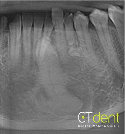
Pseudo-panoramic section
A large mixed density area is associated with the apex of the canine. It presents with multiple areas of relatively lower density with sclerotic pattern and is consistent with a focal osseous dysplasia, a self-limiting benign entity that does not require any additional assessment.
The primary canine is retained with severe root resorption and the presence of an irregular in shape high density inferior to the root situated within the thickness of the alveolar crest. This is a small compound odontoma.
The primary canine is retained with severe root resorption and the presence of an irregular in shape high density inferior to the root situated within the thickness of the alveolar crest. This is a small compound odontoma.
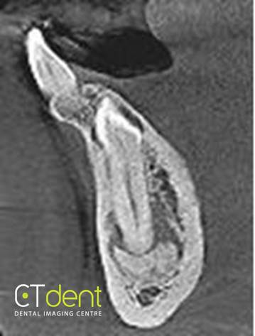
33 with focal osseous dysplasia
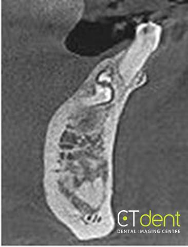
Compound odontoma inferior to the primary canine
Clinical Information: Orthodontic assessment. Ectopic LL3 positioned near midline. Retained LLC.