

Cases

Dental imaging that enhances your clinic
November 18, 2020
Case of the Month – Impacted LL3 with focal osseous dysplasia and compound odontoma
December 7, 2020Case of the Month – Radicular cyst
CBCT Scanner: KaVo OP 3D
Scanning Protocol: 5cm x 5cm FOV; 0.12 voxels
Effective Dose: 0.04 mSV
Clinical information: Upper left lateral incisor has been extracted and an implant is planned.
Click here to view and manipulate this case of the month CBCT on our Cloud Viewer
Radiographic Impression
There is no contraindication for implant placement in the area of 22. The bone is completely healed and there is good volume of bone height and width.
The following are selected images from the volume illustrating major findings
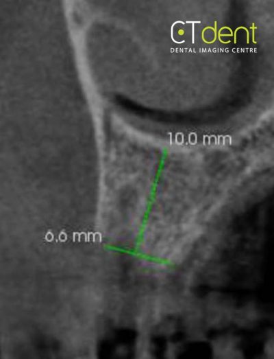
Area of the 22 with bone volume measurements
The 12 is endodontically treated with filling material extruding from the apical foramen. A periapical lesion is noted on the tooth, with material contained within. The features are suggestive of a radicular cyst.
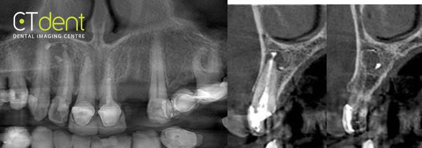
Pseudo-panoramic section Cross section of 12
Clinical information: Upper left lateral incisor has been extracted and an implant is planned.