

Cases

Using a dental imaging centre for implant CBCT scans
February 11, 2020
COVID-19 update from CT Dent
March 20, 2020Case of the month – Implant Planning in the UR5
CBCT Scanner: Instrumentarium OP300
Scanning Protocol: 8cm x 6cm FOV; 0.2 voxels
Effective Dose: 0.08 mSV
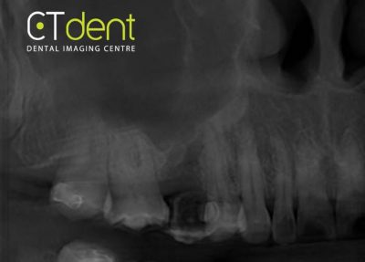
Clinical information: Implant placement planned at upper right second premolar site. Radiographic stent worn during scan.
Click here to view and manipulate this case of the month CBCT on our Cloud Viewer
Radiographic Impression
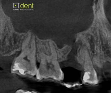
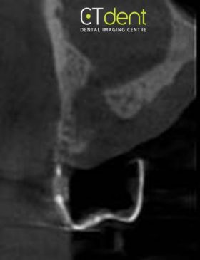
The right maxillary sinus is completely occupied by a homogenous area of increased density, and the walls appear to be sclerotic. Both findings are suggestive of a long-standing chronic sinusitis.
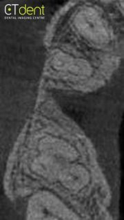
The extraction site of the maxillary right second premolar extends to include the apex of the first premolar. Clinical evaluation of the first premolar is suggested to rule out periapical inflammatory disease. There is also the appearance of a potential perforation of the cortical boundary separating the alveolar process from the maxillary sinus. This is consistent with an oral antral communication. Clinical evaluation of the site is suggested.
The following are selected images from the volume illustrating major findings
Scanning Protocol: 8cm x 6cm FOV; 0.2 voxels
Effective Dose: 0.08 mSV
Clinical information: Implant placement planned at upper right second premolar site. Radiographic stent worn during scan.
Click here to view and manipulate this case of the month CBCT on our Cloud Viewer
Radiographic Impression
The right maxillary sinus is completely occupied by a homogenous area of increased density, and the walls appear to be sclerotic. Both findings are suggestive of a long-standing chronic sinusitis.
The extraction site of the maxillary right second premolar extends to include the apex of the first premolar. Clinical evaluation of the first premolar is suggested to rule out periapical inflammatory disease. There is also the appearance of a potential perforation of the cortical boundary separating the alveolar process from the maxillary sinus. This is consistent with an oral antral communication. Clinical evaluation of the site is suggested.
The following are selected images from the volume illustrating major findings

Clinical information: Implant placement planned at upper right second premolar site. Radiographic stent worn during scan.