

Cases

Dental radiography: present and potential
July 31, 2019
CT Dent Celebrates CQC ‘GOOD’ Rating
September 10, 2019Case of the Month – The Relationship of the Lower Third Molars with the Mandibular Canal
CBCT Imaging Protocol: 16cm diameter x 6cm height, 0.2mm voxel
Effective Dose: 0.09 mSv
Clinical Information: Assessment of the relationship of the lower third molars with the mandibular canal.
Click here to view and manipulate this case of the month CBCT on our Cloud Viewer
The following are selected images from the volume illustrating major findings
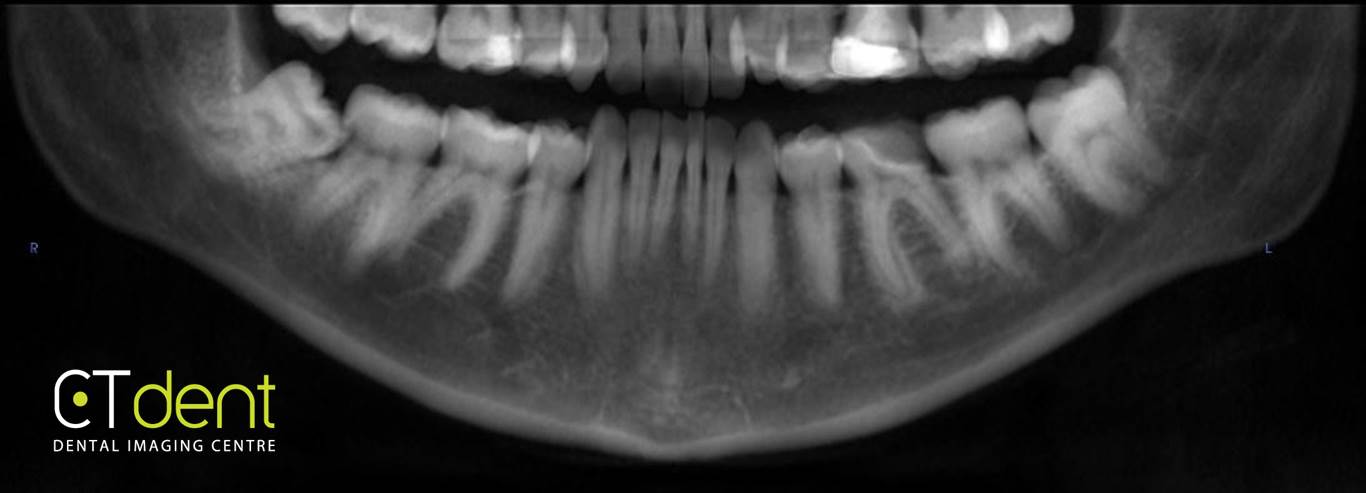
Reconstructed panoramic radiograph
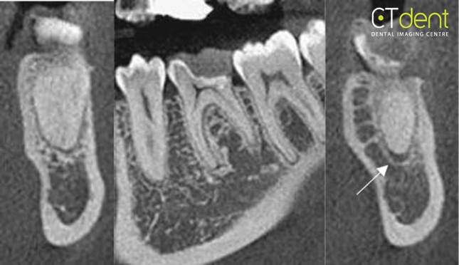
Mandibular left first molar. The PDL space surrounding the mesial root appears to be normal in width; however, the distal root apex exhibits a mild widening of the PDL space. Vitality testing of the tooth would be suggested as well as correlation of the radiographic observation with patient clinical symptoms, if any.
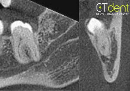
Mandibular left third molar. The mandibular canal passes in close physical proximity to the inferior surface of the root apex, without a clearly identifiable cortical separation between the apex of the root and the canal.
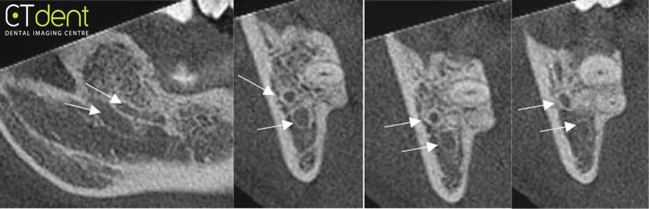
Mandibular right third molar. The mandibular canal bifurcates adjacent to the apex of the third molar with a prominent branch passing along the buccal surface of the root and the more inferior, primary canal passing inferior to the apex of the third molar. Portions of the third molar extend into, thinning and potentially perforating the lingual cortical plate.
Clinical Information: Assessment of the relationship of the lower third molars with the mandibular canal.