Cases


Case of the Month – Implant Planning at Multiple Sites in the Mandible
December 30, 2019

Using a dental imaging centre for implant CBCT scans
February 11, 2020Case of the Month – Root Treated, Symptomatic, Upper Right First Molar
CBCT Scanner: Instrumentarium OP300
CBCT Imaging Protocol: 5cm x 5cm; 0.125 voxels
Effective Dose: 0.041 mSv
Clinical Information: Root canal treatment on UR6 four months ago. Tooth is still symtomatic. Please assess for any evidence of resorption.
Click here to view and manipulate this case of the month CBCT on our Cloud Viewer
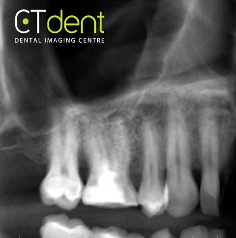
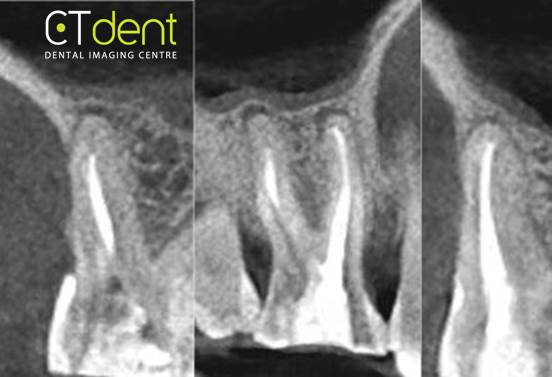
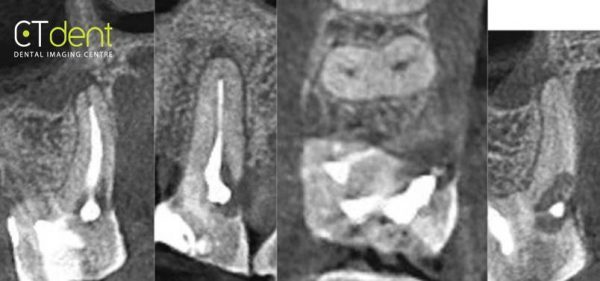
Radiographic Impression:The maxillary right first molar is root treated. There appears to be an area of resorptive change adjacent to the CEJ on the mesial lingual surface of the palatal root. Small periapical radiolucencies are also noted associated with the apices of the buccal roots, with a larger periapical radiolucency associated with the palatal root. None of the other teeth included in the volume exhibited resorptive changes.
The following are selected images from the volume illustrating major findings

Reconstructed panoramic radiograph

Distobuccal root (left) Mesiobuccal root (right)

Palatal root
Clinical information: Root canal treatment on UR6 four months ago. Tooth is still symtomatic. Please assess for any evidence of resorption.


